Z02

Z02 will extend mesoscale intracortical structural and functional imaging at 7 Tesla and include novel image acquisition, processing and modelling techniques tailored to ultra-high-resolution data to enable innovative imaging experiments in humans and primates. A worldwide unique ultra-high field MRI infrastructure will allow microstructural and functional MRI with unprecedented sensitivity and spatial resolution to extend diffusion MRI from white to grey matter and study cortical microstructure and neurofluids. Advanced structural and functional modelling of fine-grained networks in the medial temporal lobe will be developed and provided.
Principal Investigators

Dr. David Berron

Jun.-Prof. Dr. Hendrik Mattern

Prof. Dr. Oliver Speck
Co-Workers

Dr. Loris Naspi

Dr. Daniel Uher
Non-invasive imaging provides revolutionary insights
Cutting edge non-invasive human in-vivo imaging allows assessment of the brain at the meso-scale (i.e. at the level of cortical layers or neuronal ensembles). This enables knowledge transfer and mechanistic insights bridging micro-scale invasive animal research with macro-scale human brain models and interventions. Z02 and the entire CRC can leverage a worldwide unique ultra-high field MRI infrastructure with two 7T MR systems (Siemens Healthineers). Beside the state-of-the-art MAGNETOM 7T Plus (upgraded 2022), the MAGNETOM Terra.X Impulse Edition is globally the first of the newest generation of 7T scanners with a 10-times more powerful gradient system enabling microstructural and function MRI with unprecedented sensitivity and spatial resolution. This will open the window to extend diffusion MRI from white to gray matter and study cortical microstructure.
Mesoscopic Microstructural and Functional MRI
Our new 7T scanners allows us to investigate both the structure and function of the brain at a mesoscopic scale, bridging the gap between macroscale brain networks and microscopic cellular processes. We are pushing the boundaries of two key techniques: functional MRI (fMRI) and diffusion-weighted imaging (dMRI).
We are developing advanced fMRI methods to achieve sub-millimeter resolution, a level of detail that allows us to visualize brain activity in much finer detail than previously possible. This enhanced resolution will allow us to study the functional organization of the brain with greater precision, revealing how different brain regions interact and communicate during cognitive tasks. Further, we will apply sophisticated noise reduction techniques to maximize the sensitivity of our fMRI measurements, ensuring that we can detect even the subtlest changes in brain activity.
Our 7T Terra.X Impulse Edition, with its exceptionally strong gradients, allows us to acquire dMRI data with significantly improved quality and speed. This enables us to apply dMRI beyond typically performed white matter tractography and study the microstructure of the cerebral cortex. We will leverage advanced models to analyze this data, extracting crucial information about tissue properties and connectivity. This will allow us to investigate how changes in brain microstructure relate to normal brain function and to neurological disorders
Mesoscopic Imaging and Assessment of Neurofluids
Our research explores the intricate world of “neurofluids” – the various fluids and fluid-filled spaces within the brain, including blood vessels, perivascular spaces, cerebrospinal fluid, and interstitial fluid. These neurofluids play a crucial role in brain health, influencing everything from nutrient delivery and waste removal to cognitive function. We are developing and refining advanced imaging techniques to study neurofluids at a mesoscopic level.
One focus is on refining Intravoxel Incoherent Motion (IVIM) MRI, a technique that allows us to simultaneously measure blood perfusion and the movement of interstitial fluid, a potential biomarker of brain clearance. Leveraging our powerful 7T Terra.X Impulse Edition and advanced analysis methods, we strive to obtain detailed maps of these biomarkers at unprecedented detail.
We are also pushing the boundaries of structural imaging of the brain’s vasculature. Our ultra-high field MRI allows us to visualize arteries and veins with exceptional detail. We are developing and applying novel methods like vessel distance mapping to analyze the spatial relationship between blood vessels and surrounding brain tissue. This allows us to study how the organization of the vasculature influences cognition and reserve
Structural and Functional Modeling of the Human Medial Temporal Lobe
The medial temporal lobe (MTL) is a critical brain region for memory and navigation, but it’s also vulnerable to age-related changes and diseases like Alzheimer’s. Our research focuses on developing advanced methods to study the MTL’s intricate structure and function. We are developing sophisticated tools to analyze high-resolution MRI scans, enabling us to precisely map the different subregions within the MTL, including the hippocampus and extrahippocampal areas.
Our work aims to provide researchers with the resources they need to investigate the MTL. This includes developing standardized fMRI tasks and analysis pipelines for studying different types of memory, like object and scene memory. We are also providing automated tools to segment the MTL into its subregions, enabling detailed anatomical and functional studies. These tools will allow researchers to measure the volume of these subregions and track changes over time, which is crucial for understanding how the MTL is affected by aging and disease.
Beyond simply measuring volumes, we are developing new ways to analyze the fine-grained structure of the MTL, including a novel method for measuring the thickness of the hippocampus and entorhinal cortex. This will allow us to study changes at a finer scale than previously possible. Finally, we’re working to integrate data from different imaging modalities, such as structural MRI, diffusion MRI, functional MRI, and measures of neurofluids. By combining these different perspectives, we aim to gain a more complete understanding of how the MTL functions and how it changes in health and disease.
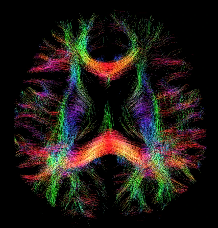
Visualization and processing by Dr. Daniel Uher.
Scanner: Siemens MAGNETOM 7T Terra.X Impulse Edition, Department Biomedical Magnetic Resonance (BMMR), Otto-von-Guericke-Universität Magdeburg
Tasks in the Collaborative Research Center
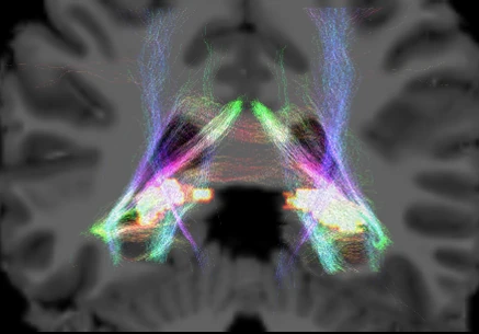
Acquisition by Dr. Yeo-Jin Yi and Dr. Yi-Hang Tung.
Visualization and processing by Dr. Daniel Uher.
Scanner: Siemens MAGNETOM 7T Terra.X Impulse Edition, Department Biomedical Magnetic Resonance (BMMR), Otto-von-Guericke-Universität Magdeburg
The aim of Z02 is the development and piloting of novel image acquisition, processing and modeling using ultra-high resolution 7 Tesla magnetic resonance imaging (7T MRI) for wider applications in human subjects and primates and thus enable innovative imaging experiments in the CRC projects. We will expand our MR imaging and analysis tools to provide a holistic assessment of neurofluids ranging from highest resolution vascular structural imaging and pattern assessment with our recently introduced vessel distance mapping (VDM) to probing the microvasculature and deriving biomarkers for perfusion and clearance. This imaging data will allow detailed structural and functional modeling of networks in the medial temporal lobe and its fine-grained subregions, including their cortical thickness cross-sectionally and longitudinally.